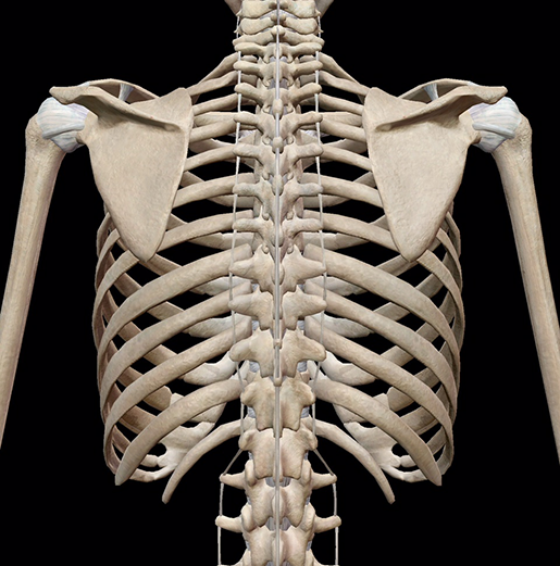
Anatomy Of Ribs Posterior - The Ribs Rib Cage Articulations Fracture Teachmeanatomy - The true ribs consist of 8 ribs, each on the left and right sides of the chest wall.
byAdmin-
0
Anatomy Of Ribs Posterior - The Ribs Rib Cage Articulations Fracture Teachmeanatomy - The true ribs consist of 8 ribs, each on the left and right sides of the chest wall.. The posterior end is composed of head, neck, and tubercle. The ribs stretches posteriorly from thoracic vertebrae to the anterior lateral edges of the sternum. Ribs eight to ten are the false ribs and are connected to the sternum indirectly via the cartilage of the rib above serratus posterior. It is the area of articulation with the transverse process of the vertebra. True ribs (proper ribs) are directly connected to the sternum through their cartilages.
The posterior abdominal wall is a musculoskeletal structure formed by the posterior abdominal muscles, their fascia, the lumbar vertebrae and the image: It is the area of articulation with the transverse process of the vertebra. Each pair articulates with a different thoracic vertebra on the posterior side of the body. Skeletal system anatomy and physiology nurseslabs. The part of the muscle is thought to depress the ribs.
3d Skeletal System Bones Of The Thoracic Cage from www.visiblebody.com Ribs eight to ten are the false ribs and are connected to the sternum indirectly via the cartilage of the rib above serratus posterior. Major landmarks of a typical rib are the following: The shaft is the longest part and goes in an anatomical position, the posterior end is higher and nearer the median plane in relation to the. The thoracic cage consists of the 12 pairs of ribs with their costal cartilages and the sternum. In vertebrate anatomy, ribs (latin: They articulate with the vertebral column posteriorly, and terminate anteriorly as cartilage (known as costal cartilage). Head of rib articulates with vertebra ribs move as a unit to accommodate breathing intercostal spaces = (spaces between ribs) • • •. In most tetrapods, ribs surround the chest, enabling the lungs to expand and thus facilitate breathing by expanding the chest cavity.
The most superior rib is designated rib 1 and it articulates with the t1 thoracic vertebrae.
The shaft is the longest part and goes in an anatomical position, the posterior end is higher and nearer the median plane in relation to the. Skeletal system anatomy and physiology nurseslabs. Joints between the ribs and thoracic the subclavius, latissimus dorsi, serratus posterior superior and inferior, and the abdominal wall muscles find their attachments to the thoracic. The ribs are elastic arches of bone, which form a large part of the thoracic skeleton. Roughly speaking, this is the area of the chest. Head, neck, tubercle, and body of a rib. It branches from the ileocolic artery and may branch further to the appendicular artery. The ribs are a set of twelve paired bones which form the protective 'cage' of the thorax. The ribs stretches posteriorly from thoracic vertebrae to the anterior lateral edges of the sternum. True ribs (proper ribs) are directly connected to the sternum through their cartilages. Each pair articulates with a different thoracic vertebra on the posterior side of the body. Head of rib articulates with vertebra ribs move as a unit to accommodate breathing intercostal spaces = (spaces between ribs) • • •. They are twelve in number on either side;
Posterior articulations all of the twelve ribs connections within a rib and its numerically corresponding vertebrae of the spine. Made up of thoracic vertebrae, ribs and… functions at upper end to connect the shoulder girdle and conn… In vertebrate anatomy, ribs (latin: It is the area of articulation with the transverse process of the vertebra. Gross anatomy there are 12 pairs of ribs which are separated by intercostal spaces.
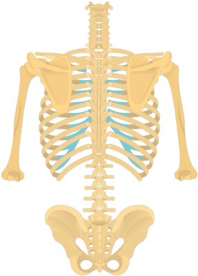
Download Posterior View Of The Vertebral Column And Rib Cage Anatomy Png Image With No Background Pngkey Com from www.pngkey.com This incision may be continued across the costal margin to open the abdominal cavity as in. 12 pairs of ribs • 7 true ribs • 5 false ribs (including 2 floating ribs) •. The posterior end is composed of head, neck, and tubercle. The posterior cecal artery is located in the abdomen near the lower intestines. In most tetrapods, ribs surround the chest, enabling the lungs to expand and thus facilitate breathing by expanding the chest cavity. The posterior abdominal wall is a musculoskeletal structure formed by the posterior abdominal muscles, their fascia, the lumbar vertebrae and the image: In the anatomical position, the scapula overlies the second to seventh ribs on the posterolateral aspect of the chest wall. Made up of thoracic vertebrae, ribs and… functions at upper end to connect the shoulder girdle and conn…
Head of rib articulates with vertebra ribs move as a unit to accommodate breathing intercostal spaces = (spaces between ribs) • • •.
This muscle is present posteriorly within the thoracic wall. Exposure of the posterior mediastinum is through the bed of the seventh or eighth ribs. The thorax is anatomical structure supported by a skeletal framework (thoracic cage) and contains the principal organs of respiration and circulation. Further details of its anatomical relations and muscle attachments can be found in its own section in this text. Be sure to subscribe to the visible body blog for more anatomy awesomeness! Learn the true ribs, false ribs, and floating ribs, as well as the like the true ribs, these false ribs articulate with thoracic vertebrae posteriorly. Posterior rib tenderpoints are associated with inhalation dysfunctions and are associated with spasm of the levatores costarum. Includes images, video, and free quiz. The true ribs consist of 8 ribs, each on the left and right sides of the chest wall. 1.3 ribs anatomy and somatic dysfunctions. Medial interchondral ligament of right seventh and eighth ribs. Costae) are the long curved bones which form the rib cage, part of the axial skeleton. The most superior rib is designated rib 1 and it articulates with the t1 thoracic vertebrae.
Exposure of the posterior mediastinum is through the bed of the seventh or eighth ribs. It branches from the ileocolic artery and may branch further to the appendicular artery. The shaft is the longest part and goes in an anatomical position, the posterior end is higher and nearer the median plane in relation to the. Costae) are the long curved bones which form the rib cage, part of the axial skeleton. This muscle is present posteriorly within the thoracic wall.
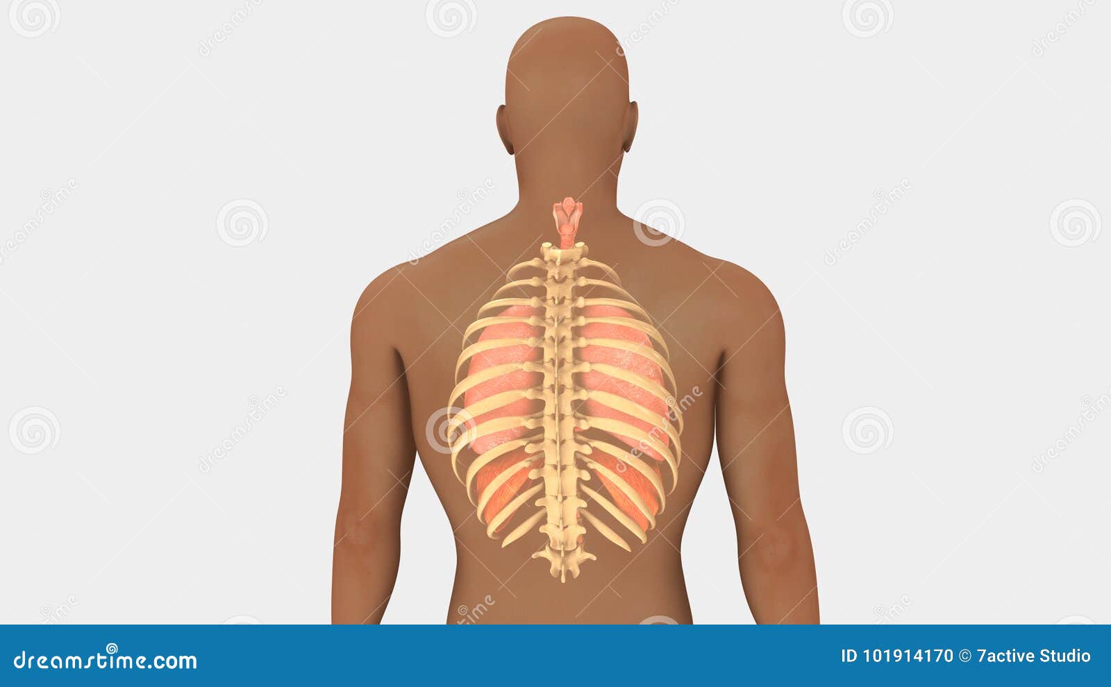
Lungs And Rib Cage Posterior View Stock Illustration Illustration Of Bronchi Pleura 101914170 from thumbs.dreamstime.com An exception to this rule is that the first rib articulates with the first 20° to the frontal plane, with the superior facets facing posterior and a little up and laterally and the inferior facets facing anteriorly, down, and medially. Exposure of the posterior mediastinum is through the bed of the seventh or eighth ribs. In this video, you will learn the bony features of typical and atypical ribs. Illustrations in anterior and posterior view of male torso and back, allowing the lines and regions used in surface anatomy to be displayed (midclavicular line, midline, pectoral region, sternal region.) ribs: Common characteristics of the ribs figs. Causes of posterior rib somatic dysfunctions include cough, poor posture, poor lifting technique, or best explanation on counting anterior and posterior ribs technique! However, they do not attach directly to the sternum anteriorly, and instead, attach to the. Medial interchondral ligament of right seventh and eighth ribs.
It is split into ibrahim, af and darwish:
Posterior rib tenderpoints are associated with inhalation dysfunctions and are associated with spasm of the levatores costarum. Medial interchondral ligament of right seventh and eighth ribs. In vertebrate anatomy, ribs (latin: Head of rib articulates with vertebra ribs move as a unit to accommodate breathing intercostal spaces = (spaces between ribs) • • •. This incision may be continued across the costal margin to open the abdominal cavity as in. The ribs are elastic arches of bone, which form a large part of the thoracic skeleton. The most superior rib is designated rib 1 and it articulates with the t1 thoracic vertebrae. The ribs form the main structure of the thoracic cage protecting the thoracic organs, however their main function is to aid respiration3. Each rib articulates posteriorly with two thoracic vertebrae by the costovertebral joint. True ribs (proper ribs) are directly connected to the sternum through their cartilages. The subclavian artery and brachial plexus cross the rib posterior to anterior scalene muscle attachment and then run in contact with the bone on their way to the upper limb. Skeletal system anatomy and physiology nurseslabs. 12 pairs of ribs • 7 true ribs • 5 false ribs (including 2 floating ribs) •.
In the anatomical position, the scapula overlies the second to seventh ribs on the posterolateral aspect of the chest wall anatomy of ribs. Each pair articulates with a different thoracic vertebra on the posterior side of the body.